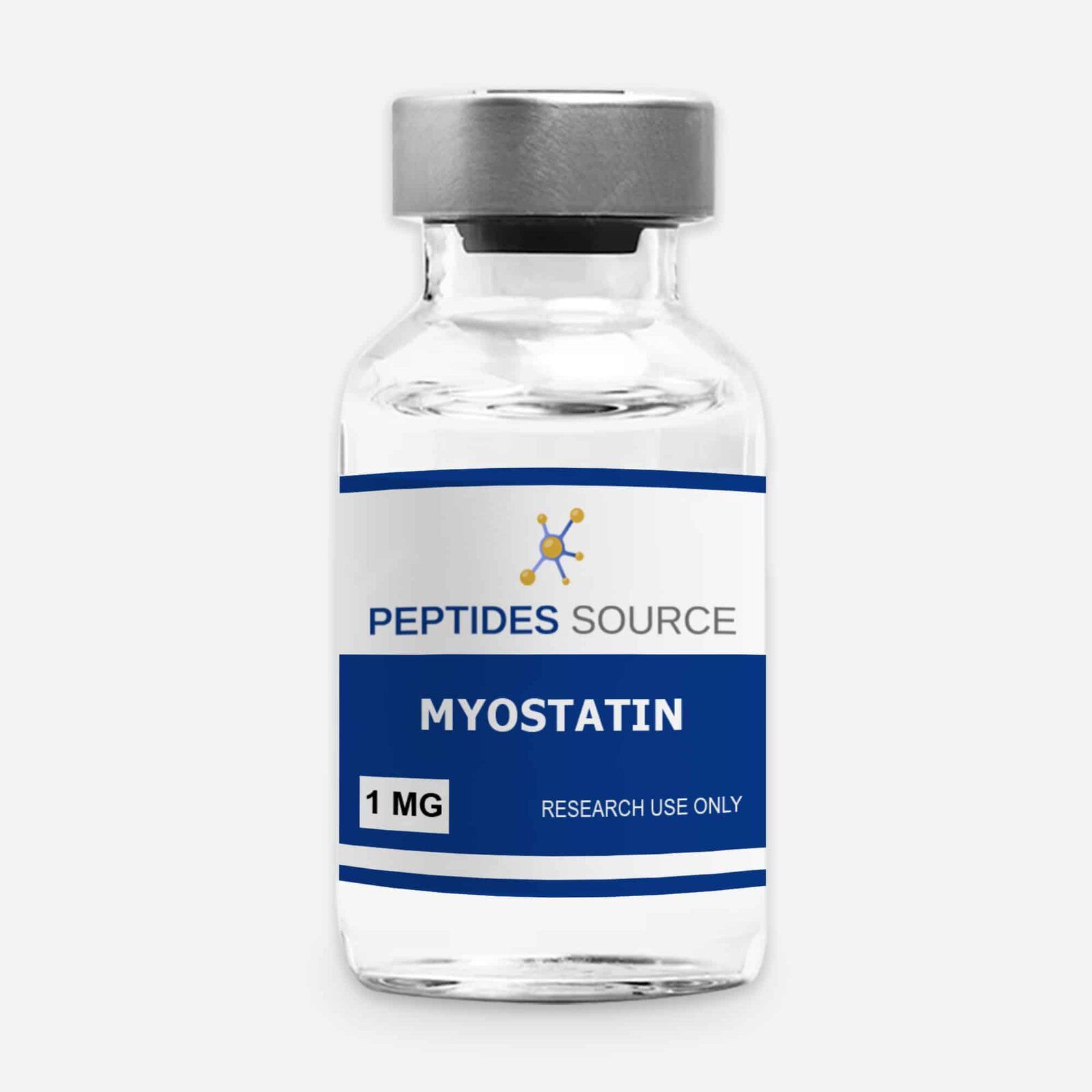
Myostatin 1mg
$15.00
You save
- Physical profile: Lyophilized powder
- This product is sold as a research chemical and not for human or animal consumption. For laboratory use by qualified professionals.
Availability: 1 in stock
Availability: Ships today if ordered and paid by 12 PM EST. (Except Saturdays & Sundays)
Product Usage
Myostatin 1mg IS INTENDED AS A RESEARCH CHEMICAL ONLY. This designation allows the use of research chemicals strictly for in vitro testing and laboratory experimentation only. All product information available on this website is for educational purposes only. Bodily introduction of any kind into humans or animals is strictly forbidden by law. This product should only be handled by licensed, qualified professionals. This product is not a drug, food, or cosmetic and may not be misbranded, misused or mislabled as a drug, food or cosmetic.
Myostatin is a naturally occurring protein belonging to the transforming growth factor-beta (TGF-β) superfamily. It is primarily responsible for regulating muscle growth by inhibiting muscle cell differentiation and proliferation. Myostatin plays a critical role in muscle homeostasis, ensuring that muscle growth does not exceed certain physiological limits.
What is Myostatin?
Myostatin, also known as growth differentiation factor 8 (GDF-8), is a protein that belongs to the transforming growth factor-beta (TGF-β) superfamily. It plays a crucial role in limiting muscle growth by inhibiting muscle cell differentiation and proliferation. In simpler terms, myostatin acts as a natural muscle growth suppressant—preventing muscles from growing beyond a certain point. This regulation is essential for maintaining balanced muscle mass, preventing excessive growth that could lead to metabolic imbalances or structural issues.How Myostatin Works (Mechanism of Action)
Myostatin is primarily produced in skeletal muscle cells and is released into the bloodstream, where it binds to the ActRIIB (activin type II receptor B) on muscle cells. This binding triggers a signaling cascade that: ⦁ Suppresses muscle cell growth (myogenesis). ⦁ Reduces protein synthesis, leading to slower muscle development. ⦁ Increases protein breakdown, causing muscle atrophy in some cases. Essentially, higher levels of myostatin = less muscle growth, while lower levels = enhanced muscle hypertrophy (growth).Myostatin structure
Myostatin is synthesized as a 375 amino acid precursor protein, which is cleaved into an active C-terminal fragment (mature myostatin).a) Full Precursor Sequence (375 Amino Acids)
MALIPVLGLLLLLLHLLGAEGPCCLEDDLLALVGAKPGEPSPGGSPCCVPAHLLQRPHTVV DGPASLSVQALRRAELRRLQAARGAAPACPAELSPPALVRASSLPLPPVPPVLRVPLSRSR ALASGGEAAAPPSLGAPATLALLLPGAVSLLAESRAYPLGSPGGSPPGGSLLLLLLGAGER AARGSRPAALLLLLLLRPALAAALAARPGAASWFSRCPRLLLPVPAHAPSSPASLAPTGRA RGATSPPQSAAPTAAPLAPPTRATRSLQDVGWLPPTQLRPPRGGRRPRRGRSRPGRALPGL EELGRLLSAELDALLSTTDSLSVTPRTPAPCCVPTRYIDFESGAQSYAESVLASSLRVPGS PVVSTLLSPGNSEPEQDDEPTPGGLAPNILSRPVILRLSVGRGLSYRGEDLAVLDDFGRLL QYVRHGEGNIFPAHAYSWAFQAKLSKLRVNQKLCNSIMDDKNADNLKKAQNLCNACCVPTK LRLDTRKSSDGSPIIPGPKVMSLQLVQRSAKTRQRYLKLKNVFNGLPNKGIQKQHSEEQQN RRKLQMWNNLMVRYNRTRQYb) Mature Myostatin (Active C-Terminal Region)
⦁ Residues: 267–375 GELVDLLQYVRHGEGNIFPAHAYSWAFQAKLSKLRVNQKLCNSIMDD KNADNLKKAQNLCNACCVPTKLRLDTRKSSDGSPIIPGPKVMSLQLV QRSAKTRQRYLKLKNVFNGLPNKGIQKQHSEEQQNRRKLQMWNNLMV RYNRTRQYKey Features:
⦁ Cysteine residues (for disulfide bond formation) ⦁ Receptor-binding sites (important for ActRIIB interaction) ⦁ Hydrophobic core regions (stabilize the dimeric structure) Molecular Formula: C₁₅₁₄H₂₄₄₈N₄₂₀O₄₃₀S₁₄ Molecular Weight: ~25 kDa (Mature Dimer) ⦁ Precursor Myostatin: ~52 kDa ⦁ Mature Active Dimer: ~25 kDa Synonyms for Myostatin (GDF-8) ⦁ Growth Differentiation Factor 8 (GDF-8) , Myostatin (MSTN),GDF8,Muscle Growth Inhibiting Factor (MGIF), Myostatin Precursor Protein , skeletal Muscle Growth Factor Pubchem substance ID: 13565157Myostatin Research
The full effects of myostatin are not entirely known. The protein, also known as growth differentiation factor-8 (GDF-8), is a member of the transforming growth factor-beta (TGF-β) superfamily and was first identified in skeletal muscle tissue. Myostatin acts as a key negative regulator of muscle growth by inhibiting muscle cell proliferation and differentiation (McPherron et al., 1997). Research has revealed that myostatin is synthesized as a precursor protein, which is cleaved to produce the active mature form that binds to activin type II receptors, initiating intracellular signaling pathways that suppress muscle growth (Rodriguez et al., 2014). Research shows that disrupting the myostatin gene in mice results in significant muscle hypertrophy, with muscle mass increasing by 200-300% compared to wild-type mice (McPherron et al., 1997). The knockout mice exhibit increased fiber size and hyperplasia, demonstrating myostatin’s crucial role in muscle mass regulation. Further studies have indicated that myostatin influences additional physiological processes, including metabolism, cardiac function, and bone density regulation (Elkasrawy & Hamrick, 2010). Attempts to inhibit myostatin activity using neutralising antibodies, receptor blockers, and gene-editing technologies have shown promising results in preclinical studies. Myostatin inhibitors have been explored as potential treatments for muscle-wasting disorders such as muscular dystrophy, sarcopenia, and cachexia (Campbell et al., 2017). Clinical trials involving myostatin inhibition in humans have demonstrated increased lean muscle mass but variable effects on strength and functionality, highlighting the need for further research to optimize therapeutic applications (Smith & Lin, 2019)⦁ Myostatin in muscle function
1. Muscle Growth and Strength
Myostatin knockout (MSTN−/−) mice exhibit a 2- to 3-fold increase in skeletal muscle mass due to both fiber hypertrophy and hyperplasia, demonstrating its powerful inhibitory effect on muscle growth (McPherron et al., 1997). Inhibiting myostatin in animal models has been associated with increased muscle fiber size, enhanced contractile strength, and improved recovery after muscle injury (Mendias et al., 2012).2. Metabolic Regulation
Beyond its effects on muscle mass, myostatin has been implicated in metabolic regulation. Myostatin-deficient mice show improved glucose uptake, insulin sensitivity, and enhanced oxidative metabolism, suggesting its potential role in metabolic disorders such as type 2 diabetes and obesity (Guo et al., 2012).3. Muscle Wasting and Disease
Elevated myostatin levels are associated with muscle atrophy in conditions like sarcopenia, cachexia, and neuromuscular diseases. Patients with Duchenne muscular dystrophy (DMD) and cancer cachexia often exhibit increased myostatin expression, contributing to muscle degeneration (Gonzalez-Cadavid & Bhasin, 2004).Clinical trials targeting myostatin inhibition using monoclonal antibodies (e.g., bimagrumab) or gene-editing techniques have shown promise in restoring muscle mass, though functional improvements remain a key research focus (Campbell et al., 2017).⦁ Fat Metabolism
1. Influence on Adipogenesis (Fat Cell Formation):
Myostatin has been observed to impact the process by which preadipocytes (precursor fat cells) differentiate into mature adipocytes:2. Effects of Myostatin Manipulation on Fat Mass:
Experimental studies manipulating myostatin levels have provided insights into its role in fat metabolism:3. Interaction with Energy Metabolism:
Beyond its direct effects on muscle and fat tissues, myostatin appears to influence overall energy metabolism:4. Therapeutic Implications:
Given its role in regulating both muscle and fat tissues, myostatin has become a target for potential therapeutic interventions aimed at treating obesity and metabolic disorders:⦁ Bone heath and development
1. Myostatin’s Role in Bone Formation and Metabolism
Recent studies have identified myostatin as a negative regulator of bone formation and metabolism. Notably, myostatin is highly expressed in fracture areas, affecting the endochondral ossification process during the early stages of fracture healing.2. Impact of Myostatin Deficiency on Bone Properties
Research involving myostatin-null mice has demonstrated that the absence of myostatin leads to increased bone formation, bone density, and bone strength. These findings suggest that myostatin inhibitors could serve as novel treatments for preventing osteoporosis.3. Therapeutic Potential of Myostatin Inhibition
The identification of myostatin as a negative regulator of muscle and bone mass has sparked significant interest in developing myostatin inhibitors as therapeutic agents for treating various musculoskeletal disorders. Inhibition of myostatin has been associated with enhanced bone mineral density and bone regeneration in animal models.4. Myostatin and Bone Regeneration
Inhibition of myostatin has been shown to accelerate bone regeneration in animal studies. Additionally, myostatin inhibition led to an increase in osteogenesis and a reduction in adipogenesis. Mice lacking myostatin exhibited less body fat and lowered adipogenesis, indicating a multifaceted role of myostatin in musculoskeletal health.5. Maternal Myostatin Deficiency and Offspring Bone Strength
Studies have demonstrated that maternal deficiency of myostatin can enhance bone biomechanical strength in adult offspring. This finding suggests that myostatin levels during development can have long-term effects on bone health.⦁ Heart function and cardiovascuar health
1. Expression of Myostatin in Cardiac Tissue
While myostatin is chiefly expressed in skeletal muscles, it is also present in cardiac muscle tissues. Under pathological conditions, such as cardiac stress or injury, the expression of myostatin in the heart is notably upregulated.2. Myostatin’s Role in Cardiac Remodeling and Heart Failure
⦁ Cardiac Remodeling: Elevated levels of myostatin have been linked to adverse cardiac remodeling, characterized by myocardial fibrosis and ventricular dilation. This remodeling can impair the heart’s pumping efficiency, potentially leading to heart failure.3. Myostatin and Cardiac Cachexia
Cardiac cachexia, a syndrome characterized by unintentional weight loss, muscle wasting, and fatigue, is commonly associated with chronic heart failure. Myostatin is implicated in this process by promoting skeletal muscle atrophy:4. Therapeutic Potential of Myostatin Inhibition
Given its involvement in adverse cardiac outcomes, myostatin presents a potential therapeutic target:5. Myostatin’s Influence on Cardiac Metabolism
Myostatin regulates energy homeostasis in the heart and prevents heart failure induced by pressure overload. It activates regulator of G-protein signaling 2, an inhibitor of Gq signaling, thereby protecting against heart failure.⦁ Classical Myostatin Signaling Pathway:
⦁ Ligand Binding: Myostatin is secreted as a latent complex and becomes active upon proteolytic cleavage. The active myostatin ligand binds to activin type II receptors (ActRIIA or ActRIIB) on the surface of muscle cells.Referenced citations
⦁ McPherron, A. C., Lawler, A. M., & Lee, S. J. (1997). Regulation of skeletal muscle mass in mice by a new TGF-beta superfamily member. Nature, 387(6628), 83-90. ⦁ Rodriguez, J., Vernus, B., & Chelh, I. (2014). Myostatin and the skeletal muscle atrophy and hypertrophy signaling pathways. Cellular and Molecular Life Sciences, 71(22), 4361-4371. ⦁ Elkasrawy, M., & Hamrick, M. W. (2010). Myostatin (GDF-8) as a key factor linking muscle mass and bone structure. Journal of Musculoskeletal and Neuronal Interactions, 10(1), 56-63. ⦁ Campbell, C., McMillan, H. J., Mah, J. K., et al. (2017). Myostatin inhibition in Duchenne muscular dystrophy: A randomized, placebo-controlled trial. Neurology, 89(16), 1751-1759. ⦁ Smith, R. C., & Lin, B. K. (2019). Myostatin inhibitors as therapies for muscle wasting associated with cancer and other diseases. Current Opinion in Supportive and Palliative Care, 13(4), 326-331. ⦁ McPherron, A. C., Lawler, A. M., & Lee, S. J. (1997). Regulation of skeletal muscle mass in mice by a new TGF-beta superfamily member. Nature, 387(6628), 83-90. ⦁ Han, H. Q., Zhou, X., Mitch, W. E., & Goldberg, A. L. (2013). Myostatin and muscle wasting: Signaling pathways and therapeutic approaches. Trends in Endocrinology & Metabolism, 24(11), 569-579. ⦁ Mendias, C. L., Bakhurin, K. I., & Faulkner, J. A. (2012). Tendons of myostatin-deficient mice are small, brittle, and hypocellular. Proceedings of the National Academy of Sciences, 109(14), 556-561. ⦁ Guo, T., Jou, W., Chanturiya, T., et al. (2012). Myostatin inhibition in muscle, but not adipose tissue, decreases fat mass and improves insulin sensitivity. PLoS One, 7(4), e33377. ⦁ Gonzalez-Cadavid, N. F., & Bhasin, S. (2004). Role of myostatin in metabolism. Current Opinion in Clinical Nutrition & Metabolic Care, 7(4), 451-457. ⦁ Campbell, C., McMillan, H. J., Mah, J. K., et al. (2017). Myostatin inhibition in Duchenne muscular dystrophy: A randomized, placebo-controlled trial. Neurology, 89(16), 1751-1759. ⦁ Rodriguez, J., Vernus, B., & Chelh, I. (2014). Myostatin and the skeletal muscle atrophy and hypertrophy signaling pathways. Cellular and Molecular Life Sciences, 71(22), 4361-4371.Protocol
Clinical Research
